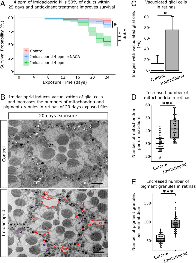
Fig. 6.
Low dose chronic exposures reduce survival and cause degeneration in the retina. (A) Survival of adults continuously exposed to 4 ppm imidacloprid. A total of 47% of adults were alive after 25 d of exposure. Flies exposed to the same dose of imidacloprid in media supplemented with 75 µg/L of NACA show 83% survival. Significance assessed with the Kaplan–Meier method and the Log-rank Mantel–Cox test (n = 100 adult flies/treatment), *P < 0.05; ***P < 0.001. (B) Electron microscopy of the retinas of flies exposed for 20 d. Dashed red lines delimitate vacuoles in glia, red arrowheads point to pigment granules, and purple arrowheads point to mitochondria. (Scale bar: 2 µm.) (C) Percentage of images that show vacuolated glial cells in the retina (10 images/adult fly; 3 adult flies/treatment). (D) Number of mitochondria per ommatidium (16 ommatidia/adult fly; 3 adult flies/treatment). (E) Number of pigment granules per ommatidium (16 ommatidia/adult fly; 3 adult flies/treatment). Shaded areas in A represent 95% confidence interval. C, D, and E, t test; *P < 0.05; ***P < 0.001.

