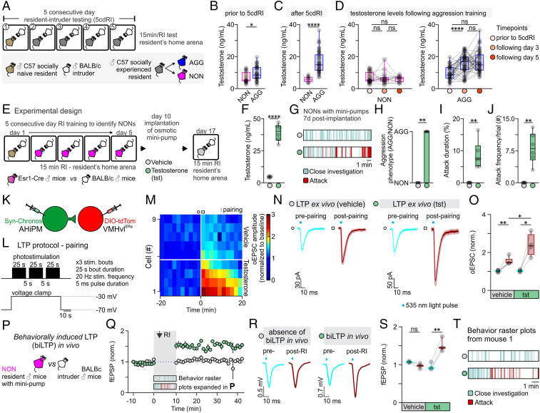
Fig. 6.
Testosterone administration leads to the expression of hypothalamic LTP and aggression in previously nonaggressive males. (A) Schematic of the experimental design used to identify aggressive (AGG) and nonaggressive (NON) males, from which tails blood samples were collected for quantification of serum testosterone levels. (B) Serum testosterone levels in NON vs. AGG mice prior to any aggression experience (n = 24 to 36 samples per group, two-sided Mann–Whitney U test, P = 0.0203 between NON and AGG groups). Mice were assigned as NON or AGG, according to whether they expressed aggression on the first day of the 5cdRI test. (C) Serum testosterone levels in NON vs. AGG mice after completion of the 5cdRI test (n = 14 to 46 samples per group, two-tailed unpaired t test, P < 0.0001 between NON and AGG groups). Mice that did not express any aggression/attack behavior throughout the 5cdRI test were assigned to the NON group. All other mice were included in the AGG group. (D, Left) Quantification of serum testosterone levels in NON mice throughout the 5cdRI test (n = 14 to 24 samples per group, Kruskal–Wallis one-way ANOVA with Dunn’s post hoc test, P > 0.9999 between “prior to 5cdRI” and “following day 3,” P > 0.9999 between “prior to 5cdRI” and “following day 5,” and P > 0.9999 between “following day 3” and “following day 5” groups). (Right) Quantification of serum testosterone levels in AGG mice throughout the 5cdRI test (n = 36 to 46 samples per group, one-way ANOVA with Tukey’s test, P < 0.0001 between “prior to 5cdRI” and “following day 3,” P < 0.0001 between “prior to 5cdRI” and “following day 5,” and P = 0.7060 between “following day 3” and “following day 5” groups). (E) Schematic of the experimental design used to identify NON mice and perform s.c. testosterone minipump implantation. (F) Serum testosterone levels in control vs. testosterone-treated mice (n = 6 mice per group, two-tailed unpaired t test, P < 0.0001 between vehicle and testosterone). (G) Representative behavior raster plots of vehicle vs. testosterone-treated mice. (H) Quantification of the number of mice that switched aggression phenotype following vehicle vs. testosterone administration (n = 0/6 in the vehicle-treated group vs. n = 6/6 in the testosterone-treated group, two-sided Mann–Whitney U test, P = 0.0022 between vehicle and testosterone). (I) Quantification of attack duration (n = 6 mice per group, two-sided Mann–Whitney U test, P = 0.0022 between vehicle and testosterone). (J) Quantification of attack frequency (number (#) of attacks per trial; n = 6 mice per group, two-sided Mann–Whitney U test, P = 0.0022 between vehicle and testosterone). (K) Schematic of the experimental design used to study the induction and modulation of LTP by testosterone in the AHiPM→VMHvl synapse in brain slices from NON mice. Note that slices were taken from animals that received T injections, but no behavioral training or other social experience. (L) Schematic of the LTP induction protocol, utilizing simultaneous photostimulation of the AHiPM terminals in VMHvl through the opsin Chronos and depolarization of the VMHvlEsr1 neuron through voltage clamp at −30 mV. (M) Heat map illustrating the magnitude of LTP induction in VMHvlEsr1 neurons from vehicle- vs. testosterone-treated mice. (N) Average current immediately prior to and following the induction of LTP in vehicle vs. testosterone conditions (light color envelope is the SE). (O) Quantification of the oEPSC, prior to and following the induction of LTP in vehicle vs. testosterone conditions (prepairing [lower 95% CI = 0.9508, higher 95% CI = 1.084] vs. postpairing [lower 95% CI = 1.274, higher 95% CI = 1.786] in vehicle conditions, n = 5 cells from three mice, two-tailed paired t test, P = 0.0038 [observed power = 0.992, Cohen’s D = 2.704, difference between means = 0.5124 ± 0.0847, 95% CI = 0.2771 to 0.7478], prepairing [lower 95% CI = 0.9349, higher 95% CI = 1.120] vs. postpairing [lower 95% CI = 1.456, higher 95% CI = 3.334] in testosterone conditions, n = 4 cells from three mice, two-tailed paired t test, P = 0.0209 [observed power = 0.889, Cohen’s D = 2.232, difference between means = 1.368 ± 0.3063, 95% CI = 0.3926 to 2.342], postpairing in vehicle [lower 95% CI = 1.274, higher 95% CI = 1.786] vs. testosterone [lower 95% CI = 1.456, higher 95% CI = 3.334] conditions, n = 4–5 cells from six mice, two-tailed unpaired t test, P = 0.0174 [observed power = 0.932, Cohen’s D = 0.6388, difference between means = 0.8652 ± 0.2974, 95% CI = 0.2044 to 1.526]). (P) Schematic of the experimental design used to trigger and record behaviorally induced LTP in vivo in NONs. (Q) fEPSP amplitude over time, prior to and following social behavior in the resident–intruder assay, in vehicle- vs. testosterone-treated NON mice (average fEPSP from n = 3 mice per group). (R) Average fEPSP amplitude immediately prior to and following the expression of social behavior in the resident–intruder assay, in vehicle- vs. testosterone-treated NON mice. (S) Quantification of fEPSP amplitude, prior to and following the induction of LTP in vehicle vs. testosterone conditions (prepairing [lower 95% CI = 1.027, higher 95% CI = 1.123] vs. postpairing [lower 95% CI = 0.7907, higher 95% CI = 1.143] in vehicle conditions, n = 3 mice, two-tailed paired t test, P = 0.1020 [observed power = 0.999, Cohen’s D = 1.6667, difference between means = 0.1081 ± 0.0374, 95% CI = −0.2692 to 0.05303], prepairing [lower 95% CI = 0.7055, higher 95% CI = 1.209] vs. postpairing [lower 95% CI = 1.027, higher 95% CI = 2.012] in testosterone conditions, n = 3 mice, two-tailed paired t test, P = 0.0098 [observed power = 0.786, Cohen’s D = 5.7787, difference between means = 0.5625 ± 0.0562, 95% CI = 0.3207 to 0.8043]). (T) Representative behavior raster plot of the same mouse treated with vehicle and 8 d after with testosterone and used for in vivo electrophysiology experiments. ns, not significant; *P < 0.05, **P < 0.01, ****P < 0.0001. In box plots, the median is represented by the center line, the interquartile range is represented by the box edges, the bottom whisker extends to the minimal value, and the top whisker extends to the maximal value. In bar graphs, data are expressed as mean ± SEM.

