Fig. 4.
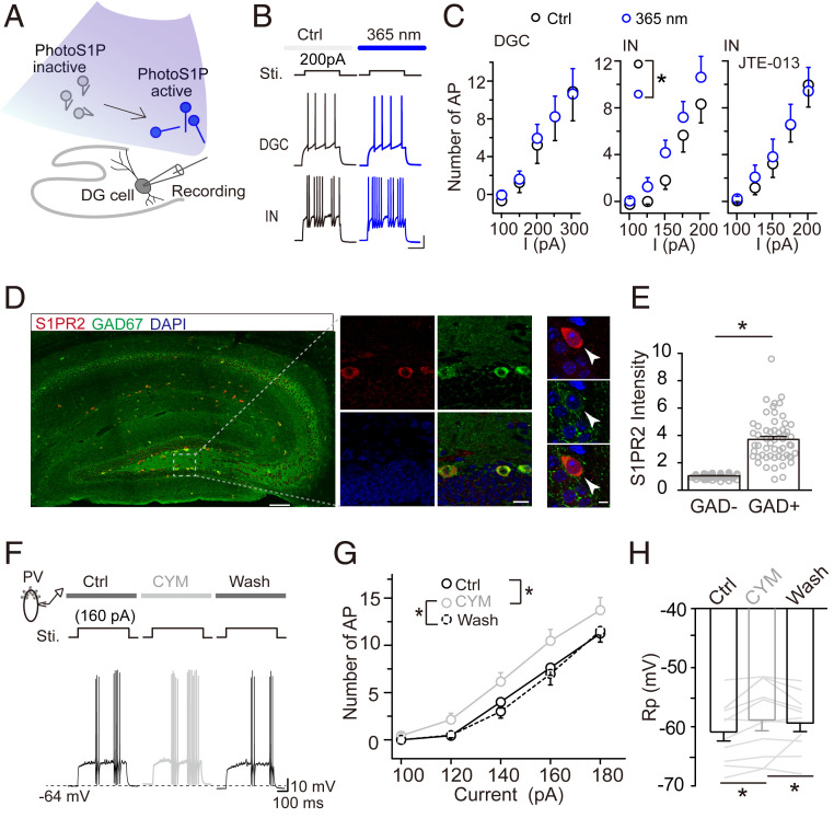
S1PR2 is specifically expressed by interneurons, and its activation causes neuronal depolarization. (A) Schematic drawing showing the switch of nonfunctional PhotoS1P (inactive) to functional (i.e., active) PhotoS1P by UV light (365 nm) illumination during slice recoding. (B) Representative images showing that active PhotoS1P increased current-evoked action potentials of interneurons (INs) but not DGCs. (Scale bar, 20 mV and 200 ms.) (C) Effect of active PhotoS1P on current-evoked action potentials (APs) of DGCs and INs (*P < 0.05, two-way RM-ANOVA). (Right) IN activity when S1PR2 was blocked by its antagonist JTE-013 (25 µM). Bicuculline (20 µM) and CNQX (10 µM) were preapplied to block inhibitory and excitatory synaptic transmission. (D and E) Preferential expression of S1PR2 in the hippocampus (arrowheads point to typical interneurons in the subgranular zone acquired by high resolution scanning) (D) and quantification in interneurons and noninterneurons in the dentate gyrus. (Scale bars are 100, 10, and 2 µm from left to right.) Normalized S1PR2 fluorescence intensity of GAD67+ vs. GAD67− cells (*P < 0.05; unpaired t test). Data were normalized to the averaged intensity of GAD67− cells (E). (F–H) Representative recordings and quantitative data showing current-evoked APs of PV+ neurons before, during, and after activation of S1PR2 by perfusion of CYM5520 (10 µM). Activation of S1PR2 significantly depolarized resting membrane potential (Rp) (*P < 0.05; two-way/one-way RM-ANOVA). (Scale bar, 10 mV and 100 ms.)

