Fig. 3.
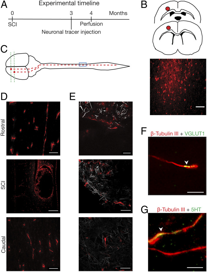
Regenerated axons invading CNF originate from the motor/sensory cortex. (A–C) Schematic drawing of the site of microinjection and tracing (red dot) and fluorescent micrograph of dextran-labeled neurons at the motor cortex (coronal section) and (D) of labeled fibers in CNF-implanted spinal cord; scales, 100 μm, 500 µm, and 100 µm, rostral, SCI, and caudal, respectively. (E) confocal micrographs of dextran-positive fibers (red) growing within the CNF (in gray, reflection mode). Scales, 20 μm, 40 µm, and 40 µm, Top, Middle and Bottom respectively. (F) and (G) Within the CNF (4 to 5 mo post SCI, different animal than above; MWCNTs are not visualized), arrowhead indicates colocalization (in yellow) of β-tubulin III-positive fibers (in red) with VGLUT1-positive puncta (in green), with 5-HT labeling (in green). Scales, 10 µm (F) and 5 µm (G).

