
Fig. 1.
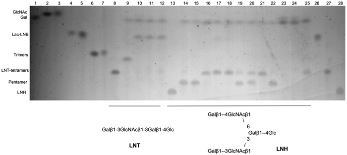
Thin layer chromatography of co-incubations of B. infantis N-acetyl-β-D-hexosaminidases with LNT or LNH after treatment with β-galactosidases. Structures are illustrated below the figure. Lanes 1–8 and 26–28: standards (as indicated in the figure); lane 9: LNT with specific β1-3 galactosidase; lanes 10–12: LNT with a β1-3 galactosidase and Blon_0459, Blon_0732 or Blon_2355. Lane 13: LNH; lanes 14–16: LNH with either a β1-3, a β1-4 or both specific galactosidases; lanes 17–19: LNH with a β1-3 galactosidase and Blon_0459, Blon_0732 and Blon_2355, respectively; lanes 20–22: LNH with a β1-4 galactosidase and either Blon_0459, Blon_0732 and Blon_2355; lanes 23–25: LNH with both β1-3 and β1-4 galactosidase, as well as either Blon_0459, Blon_0732 and Blon_2355.