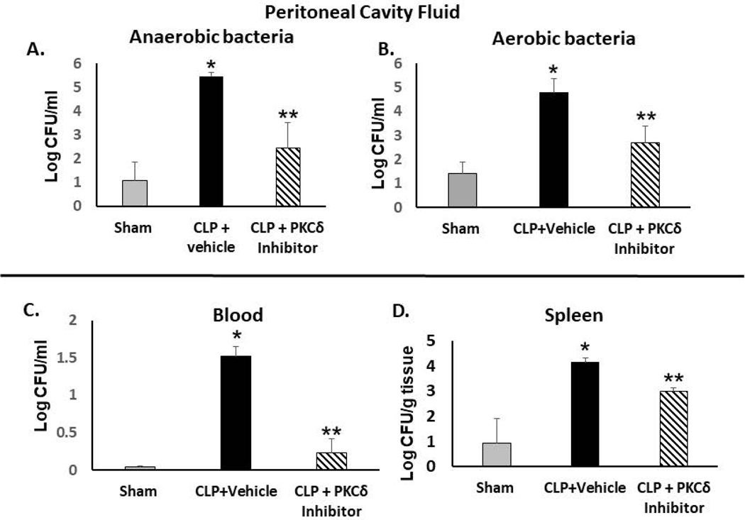
Figure 3: PKCδ inhibition improves bacterial clearance in septic rats.
Bacterial clearance was analyzed in the peritoneal cavity fluid (PCF), peripheral blood and spleens from Sham, CLP+vehicle and CLP +PKCδ inhibitor-treated rats at 24 hr post-surgery. In the peritoneal cavity fluid, bacterial colony counts were determined in serially diluted samples for 48 hrs (anaerobic, A) and 24 hr (aerobic, B). (*P< 0.01 Sham vs. CLP+ Vehicle, **p<0.05 CLP+ vehicle vs CLP+PKCδ inhibitor, n=6 rats/group). C: Peripheral blood from Sham, CLP+vehicle and CLP +PKCδ inhibitor treated rats at 24 hr post-surgery (*P< 0.001 Sham vs. CLP+ Vehicle, **p<0.001 CLP+vehicle vs CLP +PKCδ inhibitor, n=6 rats/group). D: Spleens collected from Sham, CLP+ vehicle and CLP +PKCδ inhibitor rats (*P< 0.05 Sham vs. CLP+ Vehicle, **p<0.05 CLP+ vehicle vs CLP+PKCδ inhibitor, n=3 rats/group. Bacterial spreading was quantified as CFU (colony-forming unit) per ml of fluid (PCF and blood) or as CFU/g tissue (spleens).

