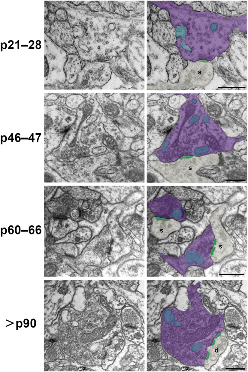
Figure 1.
Development of excitatory SBB morphology. Spinules (blue) were observed within cortical boutons (purple) at every postnatal day age group examined. In 2D TEM sections, spinules were occasionally observed invaginating into SBB profiles (e.g., p21–p28); however, most spinules appeared as ovoid or circular double membrane-bound structures encapsulated within their “host” bouton. PSDs (green) at SBB synapses sometimes contained perforations (e.g., p60–p66 and >p90 panels). Note that SBBs at times displayed multiple spinule cross-sections (i.e., 2D profiles of putative spinules), yet in our 2D TEM analyses, it was not possible to attribute these to single spinules with complex morphologies, or to multiple spinules protruding into a single SBB. s = postsynaptic spine; d = postsynaptic dendrite. Scale bars = 0.5 μm.

