Figure 4.
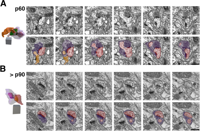
FIBSEM images of SBBs in p60 and >p90 V1. A, Adjacent FIBSEM images of an SBB in L4 of V1 from a p60 ferret. FIBSEM images are ∼25 nm apart in z (depth) on average, picked to display the progression of spinule invagination and engulfment into the SBB. Top panel, Raw FIBSEM images. Bottom panel, Pseudo-colored to highlight SBB (purple), adjacent axon/bouton (orange), and postsynaptic spine (red). Note the adjacent bouton (orange) with a synapse onto a spine that protrudes a spinule into this SBB at the bottom left of the first image in the series, and the spinule from the postsynaptic spine that invaginates into this SBB across the middle of its perforated PSD. Left, Full reconstruction of this SBB showing transparent bouton (purple) with engulfed postsynaptic spine (red) and adjacent bouton (orange) spinules. Note the horseshoe-shaped perforated PSD. Identical SBB as shown in Figure 6C,C1. B, Adjacent FIBSEM images of an SBB in L4 of V1 from a >p90 ferret. Top and bottom panels arranged and colored as in A. Note the postsynaptic spine (red) that sends its spinule into its SBB (purple) partner from the edge of the PSD. Left, Full reconstruction of this SBB (purple), made transparent to show the engulfed anchor-like spinule from its postsynaptic spine partner. Macular-shaped PSD (green) appears yellow within spine. Identical SBB as shown in Figure 7D,D1. 2D scale bars = 0.5 μm; 3D scale cubes = 0.5 μm3.

