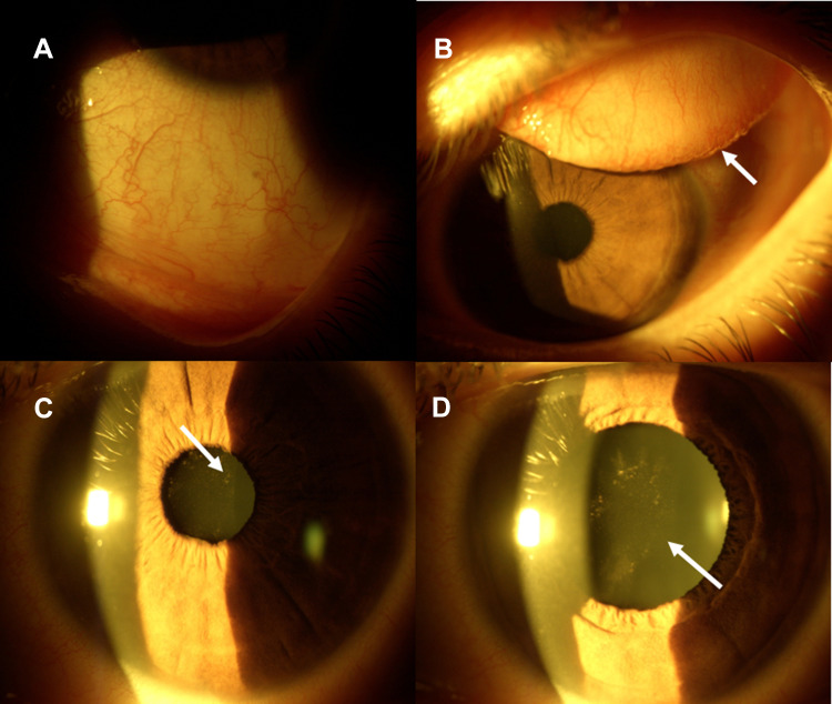
Figure 1.
SARS-CoV-2 Anterior Uveitis. Conjunctival diffuse hyperaemia (A) Acute Follicular conjunctivitis with sub-tarsal follicular hypertrophy ((B) white arrow). Mild miosis with pigmentary acute anterior uveitis ((C) white arrow). After pharmacological mydriasis, pigmentary and whitish inflammatory precipitates on the anterior capsule of the crystalline lens with initial lens opacity were evident ((D) white arrow).

