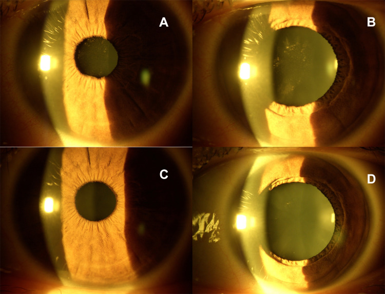
Figure 2.
SARS-CoV-2 Anterior Uveitis. Mild miosis with pigmentary acute anterior uveitis (A) before treatment. Pigmentary and inflammatory whitish precipitates on the anterior capsule of the crystalline lens with initial lens opacity (B) after pupil dilatation, before the therapy. Biomicroscopic evidence of cleared anterior capsule of the crystalline lens in miosis (C) and in mydriasis (D) with the disappearance of pigmentary and diffuse whitish inflammatory deposits after combined topical therapy.

