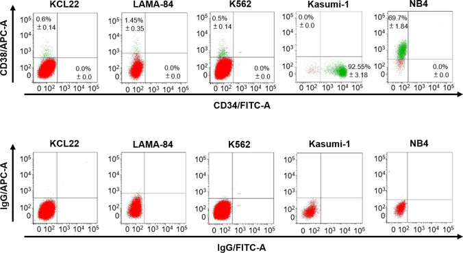
Fig. 2.
Evaluation of the expression of the CD34 and CD38 cell surface markers in CML cell lines. The expression of CD34 and CD38 in routinely cultured cells was assessed by flow cytometry. Dot plots from one representative experiment are shown. The background signal was measured by cell staining with matched-isotype IgG controls. Values represent mean ± SD of data from three independent experiments. Kasumi-1 and NB4 cells were used as positive controls for CD34 and CD38, respectively

