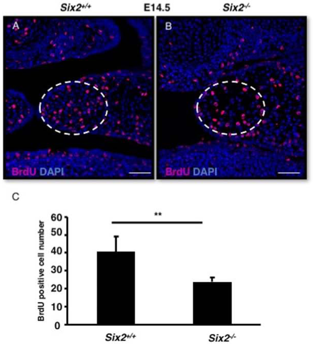
Fig. 5. Six2−/− embryos have decreased mesenchymal cell proliferation in palate shelves at E14.5.

A. In order to label dividing cells, BrdU was injected into an E14.5 stage pregnant female and embryos were harvested 2 hours later. Immunostaining was used to label BrdU+ cells on the palate shelves of WT and Six2−/− embryos. C. The number of proliferative cells found in E14.5 WT and Six2−/− palate mesenchyme were counted, and a statistically significant decrease in proliferation was observed in the Six2−/−. Scale Bars, 50 μm. Error bars show SEM. **: p-values<0.01.
