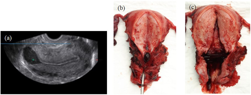
Figure 2.
Accumulation of fluid/blood in the niche. (a) Image of niche using transvaginal ultrasound in mid-sagittal plane with intrauterine fluid accumulation in the large niche. (b and c) Macroscopic image of a uterus with a niche, removed by laparoscopy because of abnormal uterine bleeding and dysmenorrhoea. Clear accumulation of mucus and blood in the niche can be recognized.

