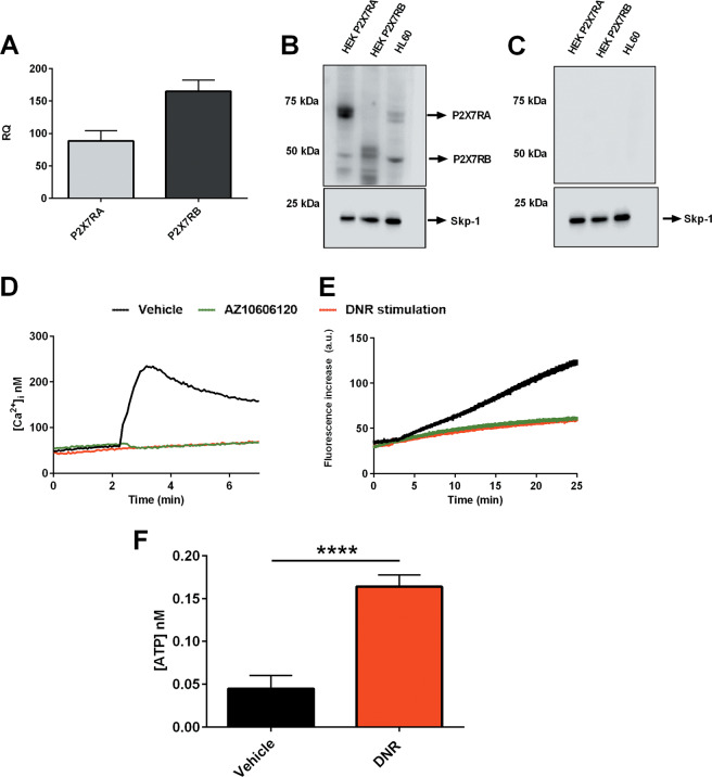
Fig. 5. HL-60 human promyelocytic cell line expresses P2X7RA and B isoforms and releases ATP upon DNR treatment.
a mRNA expression of P2X7RA (light gray) and P2X7RB (dark gray) in HL-60 cells (n = 6). b P2X7R isoforms expression revealed with an antibody directed against P2X7R extracellular domain, P2X7RA corresponds to a protein of ~70 KDa, while P2X7RB corresponds to a protein of ~50 KDa. Lane 1: HEK-293 transfected with P2X7RA, lane 2: HEK-293 transfected with P2X7RB, and lane 3: HL-60. Gel loading control Skp-1 (~20 kDa). c Immunoblot obtained with extracellular-domain directed anti-P2X7R antibody preincubated with blocking peptide (1/2 ratio, see “Materials, subjects, and methods”). Lane 1: HEK-293 transfected with P2X7RA, lane 2: HEK-293 transfected with P2X7RB, and lane 3: HL-60. Gel loading control Skp-1. d Representative traces showing an increment of intracellular calcium following stimulation with 500 µM BzATP alone (black) or applied after 5 min pretreatment with AZ10606120 (green) or following stimulation with 200 nM DNR alone (red). e Representative traces showing ethidium bromide uptake following stimulation with 500 µM BzATP alone (black) or applied after 5 min pretreatment with AZ10606120 (green) or following stimulation with 200 nM DNR alone (red). f Extracellular ATP (nM) was measured in the culture supernatants as described in “Materials, subjects, and methods.” HL-60 cells were plated at 5 × 10⁵ cells per ml and treated for 24 h with vehicle (PBS, black) and DNR (200 nM, red) (n = 8 for each group, vehicle versus DNR ****P < 0.0001).

