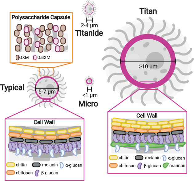
Figure 3. Cell surface alterations and cell size changes in C. neoformans.
Model illustrating the cell surface alterations and cell size phenotypes in C. neoformans. The outside of the C. neoformans cell consists of a cell wall (highlighted in pink) and polysaccharide capsule. Typical cells (5–7 μm in diameter) have a capsule predominantly composed of GXM and GalXM (orange box). The cell wall of typical cells contains chitin, chitosan, α-glucan, melanin, and β-glucan (left pink box). Titan cells (>10 μm in diameter) exhibit a thickened cell wall with an increase in chitin, decreased glucan, and a layer of mannan (right pink box) [59]. Micro cells (<1 μm in diameter) also display thickened cell walls [66]. Titanides are 2–4 μm in diameter and are oval in shape. The cell wall of titanides are thinner than typical cells [71]. Abbreviations: GXM, glucuronoxylomannan; GXMGal, glucuronoxylomannogalactin.

