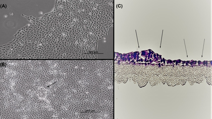
Figure 1. Morphology of 16HBE cell cultures.
Phase contrast image of a subconfluent (A) and 1 day post-confluent (B) cell layer grown in a Falcon T75 culture flask as described in ‘Materials and methods’ section (100×). A dome/hemicyst is pointed out by the black arrow. (C) A 16HBE confluent cell layer on a permeable polycarbonate filter was cross-sectioned and then stained with Hematoxylin and Eosin, showing that 16HBE can form both simple monolayer (dashed arrows) and multilayered (solid arrows) epithelial layers at confluence.

