
Figure 3.
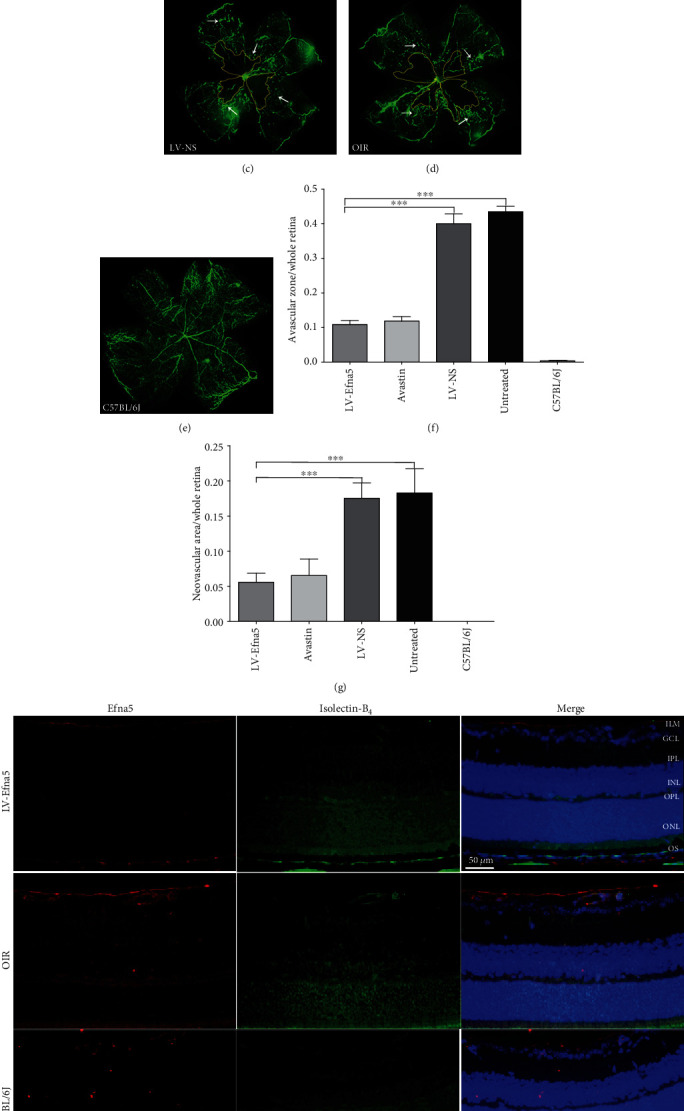
Intravitreal injection of LV-Efna5-shRNA markedly reduced the avascular area and neovascularization in the retinas. (a–e) After receiving intravitreal injections, mice from each group were perfused with fluorescein isothiocyanate- (FITC-) dextran (green) at P17; retinal flat mounts were prepared for each sample and analyzed by fluorescence microscopy. (f) Quantification of avascular areas in the retinas of OIR mice. Avascular areas were featured by a lack of fluorescence. (g) Quantification of the neovascular area in the retinas of OIR mice. Neovascularization areas were featured by hyperfluorescence. n = 5 animals per group. Scale bar represents 500 μm; area enclosed by yellow line: avascular area; white arrow: neovascular area; ∗∗∗P < 0.001. (h) Retinas of mice with indicated treatment were harvested at P17. Immunofluorescence staining was performed with anti-Efna5 antibody (red) and Isolectin-B4 (green). Scale bars represent 50 μm; ILM: internal limiting membrane; GCL: ganglion cell layer; IPL: inner plexiform layer; INL: inner nuclear layer; OPL: outer plexiform layer; ONL: outer nuclear layer; OS: outer segment.