
Fig. 8.
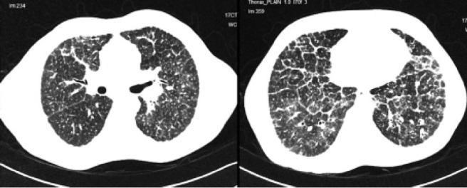
Composite image of the HRCT image of Patient 4 showing (left, upper lobes) random nodules with perilymphatic predominance, beading of the right oblique fissure and perivascular nodules and (right, lower lobes) marked interlobular septal thickening in the lower lobes associated with random nodules