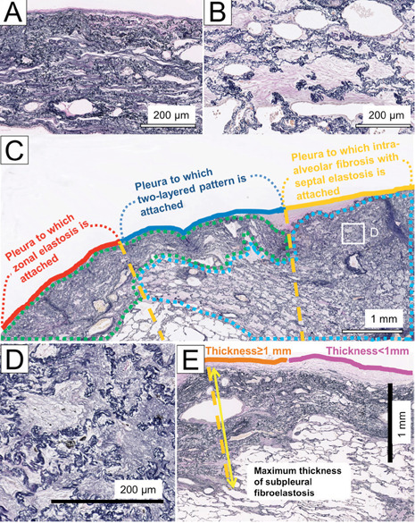
Fig. 1.

Elastica van Gieson-stained sections in a patient with IPPFE. (A) Zonal elastosis. (B) Intra-alveolar fibrosis with septal elastosis (IAFE). Fibroelastosis in PPFE was segmented into zonal elastosis (green lesion) or IAFE (blue lesion) (C). We then determined the part of the pleura associated with zonal elastosis, IAFE, or a two-layered pattern involving zonal elastosis and IAFE. Each fibroelastosis pattern was decided on the orange-dotted vertical lines originating at the pleura and extending inward from the pleura. In this example, the percentages of the pleural length with zonal elastosis, a two-layered pattern, and IAFE were calculated to be 32.9%, 38.4%, and 28.8%, respectively. Fine elastic fibres are sometimes observed in old collagen-filled alveoli with septal elastosis (D, inset of C). (E) Maximal thickness of the subpleural fibroelastosis and pleura with subpleural fibroelastosis <1 mm or ≥1 mm thick
