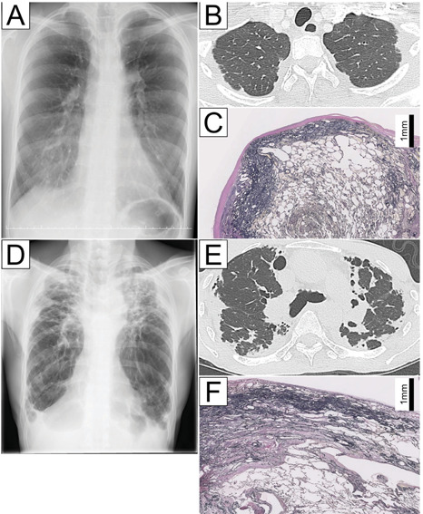
Fig. 2.

Case 1 (A-C). Chest radiography and computed tomography findings for a 23-year-old female with IPPFE demonstrating modest wedge-shaped alveolar consolidation in the subpleural region of the upper lobes (A and B). An Elastica van Gieson-stained section of the upper lobe showing a zonal elastotic lesion (C). Case 2 (D-F). Chest radiography and computed tomography findings for a 64-year-old man with IPPFE demonstrating extensive wedge-shaped alveolar consolidation in the subpleural region of the upper lobes (D and E). An Elastica van Gieson-stained section of the upper lobe showing a two-layered fibroelastosis pattern (F).
