
Figure 2.
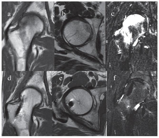
Magnetic resonance imaging (MRI) before and after micrografts treatment. The femoral head MRI was performed using a standard technique for artefact suppression. A complete regression of the necrotic area accomplished by restoring of osseous structure suggesting an autologous micrografts-induced bone regeneration