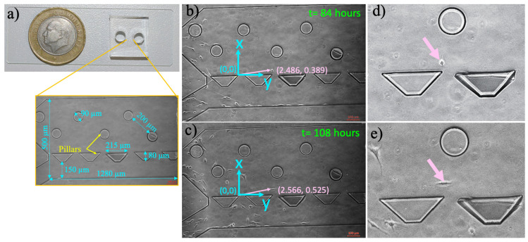
Figure 1.
Microfluidic cell culture platform and measurement of single-cell migration in the microfluidic device. (a) The polydimethylsiloxane (PDMS) microfluidic chamber on a glass slide, (b) the micrographs of microfluidic chamber with pointed pillars and dimensions. (c) The blue line indicates the position of the coordinate system (0, 0) with x- and y-axis. The pink arrow points to the position of a cell according to origin, (b) x: 2.486, y: 0389 at 84 h, (c) x: 2.566, y: 0.525 at 108 h, (d) and (e) demonstrate the zoomed images of this cells in (b) and (c), respectively. The scale bar shows 100 µm.

