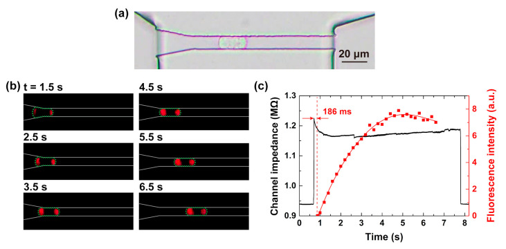
Figure 4.
(a) Microscopic image of a single A549 cell inside a constriction channel observed in the bright field, (b) time-lapse fluorescent images of the propidium iodide (PI) delivery during cell passing with a voltage of 3 V. (c) The correspondences of the channel impedance (black line) and fluorescence intensity (red dot-line) over time when the cell in (b) passed through the channel.

