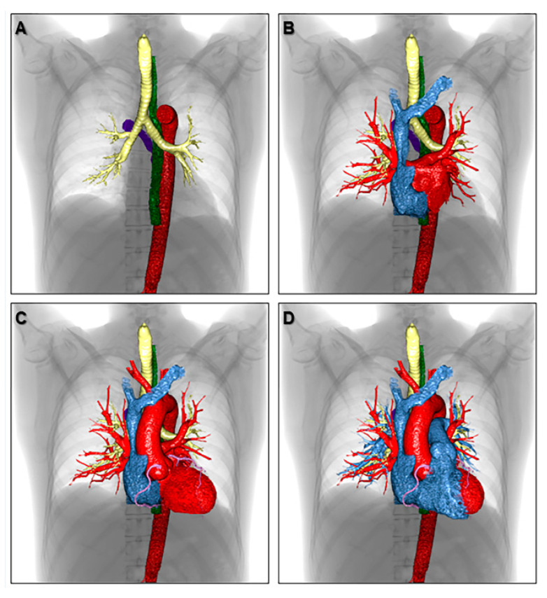
Figure 8.
Volume-rendered images showing the three-dimensional relationship of each anatomical structure in the thorax. The azygos vein (purple), descending aorta (red), esophagus (green), trachea, and bronchi (yellow) are shown in panel (A). Both atria are added in panel (B). The left ventricle, aortic root, ascending aorta, aortic arch, and coronary artery are added in panel (C). The right ventricle, pulmonary root, pulmonary trunk, and pulmonary arteries are reconstructed in panel (D) to finalize the components of cardiac silhouette.

