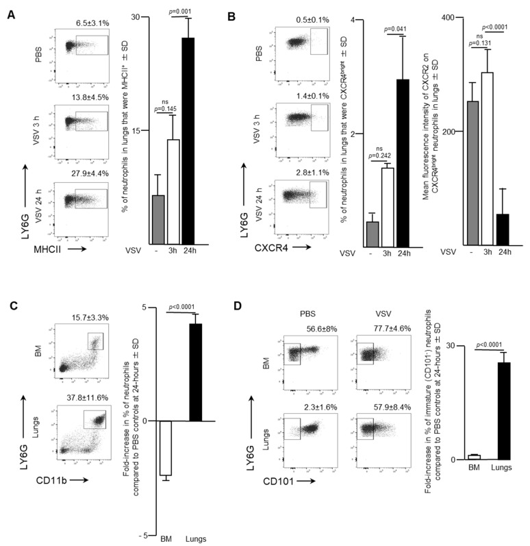
Figure 3.
Neutrophils accumulated in the lungs of mice following intravenous administration of vesicular stomatitis virus (VSV). Six- to eight-week-old female C57BL/6 mice were injected intravenously with 1 × 10⁹ plaque-forming units of VSV or phosphate-buffed saline (PBS) and euthanized three- or 24-h (h) later. Femur-derived bone marrow (BM) and lungs that had been purged of blood were used to quantify neutrophils by flow cytometry. Representative dot plots and graphs with means and standard deviations (SD) (n = 8/group) are shown for the percentage of (A) pulmonary neutrophils (defined as CD45+Ly6GhiCD11bhi) that expressed major histocompatibility complex class II (MHCII), (B) pulmonary neutrophils that were CXCR4bright (left graph) and the mean fluorescence intensity of CXCR2 on these CXCR4bright neutrophils (right graph), and (C,D) the fold-change in the percentage of; (C) neutrophils; or (D) immature neutrophils (CD101-) in the BM and lungs compared to PBS-treated control mice. (ns = not significantly different).

