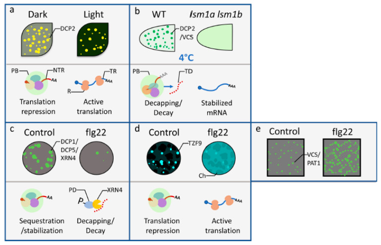
Figure 2.
P-body dynamics and functions in mRNA decay and translation repression in response to abiotic or biotic environmental cues. (a) P-bodies and associated global translation repression in Arabidopsis cotyledons decreased during transition from dark to 4-h light treatment [37]. (b) Low temperature induced formation of p-bodies and specific mRNA decay in Arabidopsis root tips was dependent on LSM1A and LSM1B [40]. (c) Yu et al. (2019) demonstrated that the treatment of flg22 triggered the disassembly of DCP1-associated p-bodies and differential mRNA decay in Arabidopsis protoplasts [42]. (d) The treatment of flg22 led to the decrease of TZF9-containing granules and the release of translation repression in Arabidopsis protoplasts [43]. The spherical dark-shade clusters in the background of the upper panel of (d) are chloroplasts (Ch). Note that the TZF9 fluorescence signal changed from distinct granules to a diffusive cytoplasmic pattern. (e) In contrast to (c), the treatment of flg22 induced the formation of PAT1-associated p-bodies in Arabidopsis root epidermal cells [44]. For each subfigure, the upper panel illustrates the localization of SGs within the respective cells/tissues under mock (control) or treatment (light, flg22) conditions. The lower panels are graphical representations of mRNA destinies correlating with the functions of p-bodies. DCP2: DCP2-YFP focus; DCP2/VCS: GFP-DCP2 or GFP-VCS focus; DCP1/DCP5/XRN4: DCP1-GFP, DCP5-GFP, or XRN4-GFP focus; TZF9: TZF9-GFP focus; VCS/PAT1: VCS-GFP or PAT1-GFP focus; PB: p-body; NTR: non-translating mRNA associated with PB; R: ribosome; TR: translating mRNA; TD: target of decapping machinery; P: phosphate; PD: phosphorylated DCP1.

