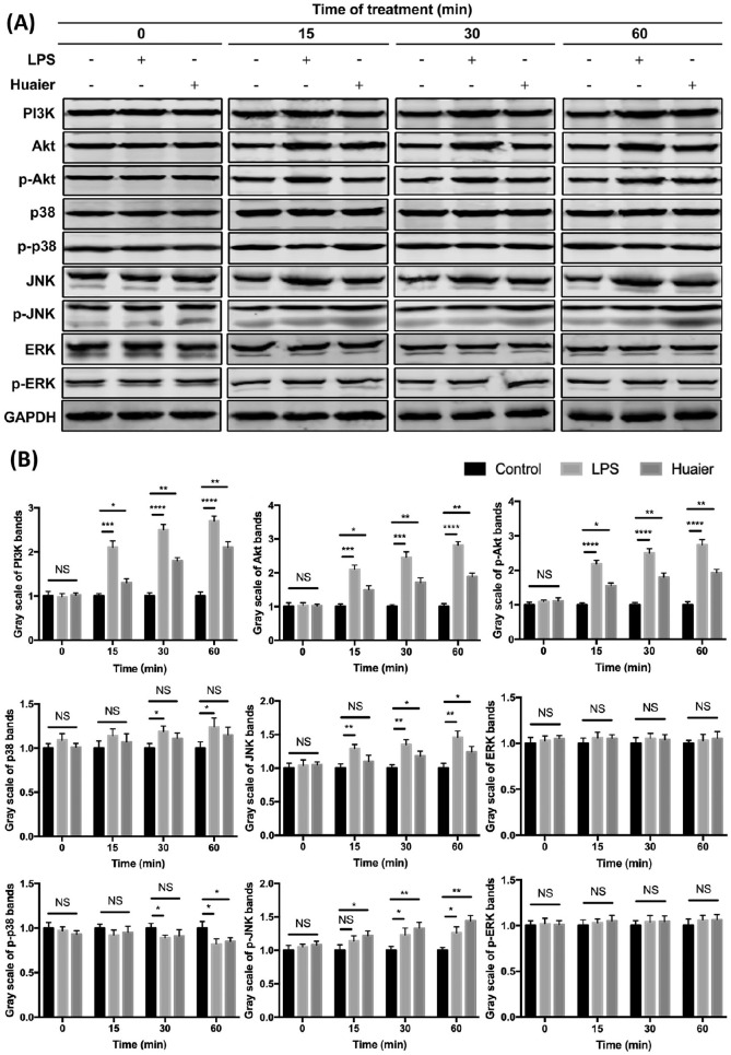
Figure 7.
Western blot analyses of signaling proteins in BMDCs from different groups. BMDCs were treated with 1 µg/ml LPS or 4 mg/ml Huaier extractum for 0, 15, 30, and 60 minutes. PI3K, Akt, p-Akt, p38, p-p38, JNK, p-JNK, ERK, p-ERK, and GAPDH expressions in treated BMDCs were analyzed by Western blot (A). Gray scale analyses of different protein bands were shown (B).
*P < .05, **P < .01, ***P < .001, ****P < .0001.

