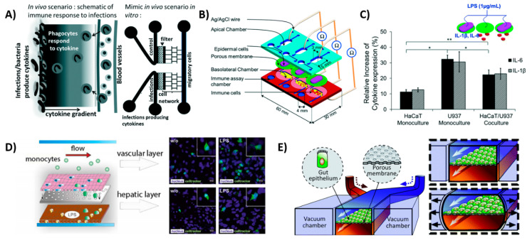
Figure 4.
Schematic views of OoCs with immune cell components. (A) Inflammation-on-a-chip by Gopalakrishnan et al. [75] mimicking the cytokine gradient in a normal wound and the migration of immune cells. Reproduced from Ref. [75] with permission from The Royal Society of Chemistry. (B) skin-on-a-chip by Ramadan et al. [80] with dermal and epidermal layer, showing the interactions of DCs with the keratinocytes and (C) showing the expression of inflammatory cytokines IL-6 and IL-1β in the device with a keratinocyte (HaCaT) monoculture and a dendritic cell (U937) monoculture and the co-culture. Reproduced from Ref. [80] with permission from The Royal Society of Chemistry; (D) liver-on-a-chip by Gröger et al. [89]; the top layer consisting of endothelial cells and macrophages and the bottom layer consisting of hepatocytes and hepatic stellate cells. LPS is used to induce an inflammatory response and monocytes start migrating; (E) gut-on-a-chip by Kim et al. [90], showing the peristaltic motion as a result of the two vacuum chambers. This chip model is used to incorporate immune cells in later stages of the research [82]. Reproduced from Ref. [82] with permission from The Royal Society of Chemistry.

