Figure 4:

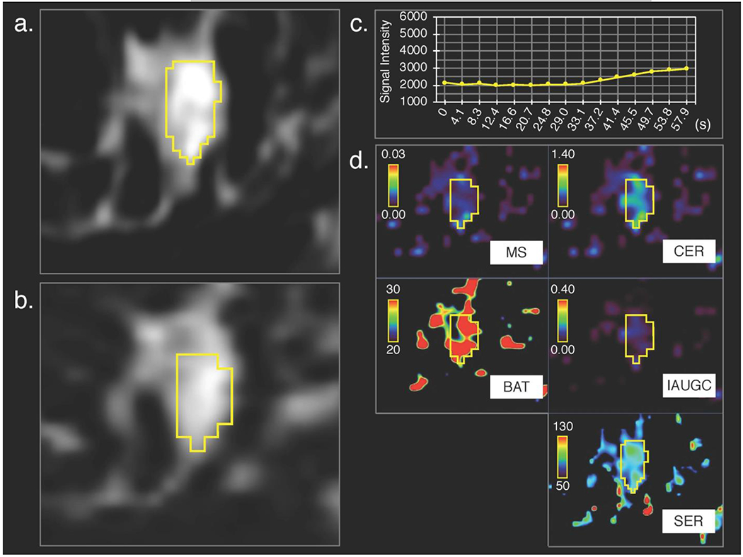
Fibroadenoma in left breast of a 50-year-old woman, (a) 3D volumetric segmentation (yellow frame) was performed on the 1st phase of conventional dynamic contrast-enhanced magnetic resonance (DCE-MR) images, (b) cloned to all the other DCE-MRI phases; the 15th ultrafast DCE MR image is shown as a representative. Compared with invasive lobular carcinoma shown in Figure 3, (c) time–signal intensity (average of the whole lesion) curve of ultrafast DCE-MRI show slower increase of signal intensity and (d) parametric maps show less difference between the lesion and surrounding tissue.
