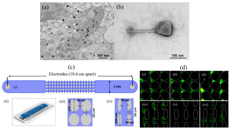
Figure 5.
Virion enrichment with iDEP traps [104]. (a) Transmission electron microscope (TEM) image of Salmonella (SPN3US-infected). The SPN3US progeny particles are indicated with white arrowheads. (b) Image of a single SPN3US (Negatively stained) virion showing head (containing dsDNA genome) and tail. (c) Sketch and 2D/3D design of iDEP channel. (i): Top view showing schematic of a full channel. (ii): 3D representation of the channel. (iii): Circle diameter 200 µm with gap of 20 µm, (iv): Oval shape posts. (d) Circle posts: (i) SPN3US virions at 1200 V, (ii) P. aeruginosa phage ϕKZ at 1100 V, and (iii) 1100 V applied on P. chlororaphis phage 201ϕ2-1. Oval posts: (iv) SPN3US virion at 800 V, (v) P. aeruginosa phage ϕKZ at 750 V and (vi) P. chlororaphis phage 201ϕ2-1 at 750 V. [104]. ©Reprinted (adapted) with an open access article distributed under the Creative Commons Attribution License which permits unrestricted use, distribution, and reproduction in any medium.

