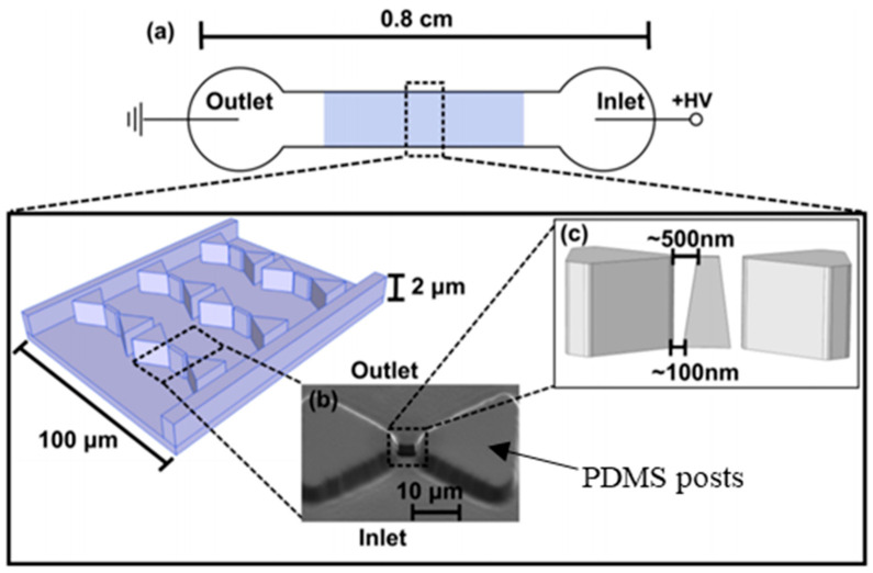
Figure 8.
Schematic top view of the iDEP micro device (not to scale) [116]. (a) An electric potential difference applied across microchannel inlet and outlet showing insulated the array of posts at the center of the device. (b) A tiny nanoconstriction outlet where nm-size proteins are manipulated between the tips of triangular microposts. (c) Image showing triangular post dimensions along with gap between nanoconstrictions in the PDMS mold [116]. ©Figure 8 adapted with Copywrite Clearance and reprinted (adapted) with permission from: Copywrite (2001) Royal Society of Chemistry, ID 1048996-1.

