Figure 5.

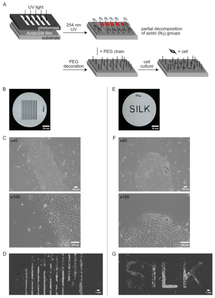
Spatial patterning of NIH3T3 fibroblasts on AzidoSilk film modified with DBCO-mPEG of 20 kDa chain length after partial irradiation of 254 nm UV light through patterned photomasks. Fibroblasts were cultured for 3 days in Eagle MEM supplemented with 10% FBS. (A) Experimental procedure. UV light was first irradiated to AzidoSilk film through a photomask for the partial decomposition of azido groups. After the PEG chain decoration, cells were cultured on the AzidoSilk film. (B,E) Photomasks used in this study. Scale bars are 10 mm. (C,F) Fibroblasts on the boundary of UV-irradiated and nonirradiated areas on the third day of culturing. Scale bars are 100 μm. (D,G) Wide views showing patterned adhesion of fibroblasts. Culture medium was removed, and cells were immobilized by 100% methanol followed by drying. Cells turned into white after fixation. Scale bars are 1 mm.
