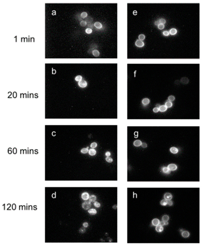
Figure 6.
Endocytosis of [K7(NBD),Nle12]α-factor detected with fluorescence microscopy. Cells expressing full-length receptors (left panels) or truncated receptors (right panels) from multicopy plasmids were incubated with the fluorescent ligand at 30 °C for (a,e) 1, (b,f) 20, (c,g) 60, and (d,h) 120 min. The ranges of image intensities in panels (e–h) are approximately 2-fold greater than those for panels (a–d), indicative of the stronger fluorescence from the truncated receptors. Taken with permission from [70].

