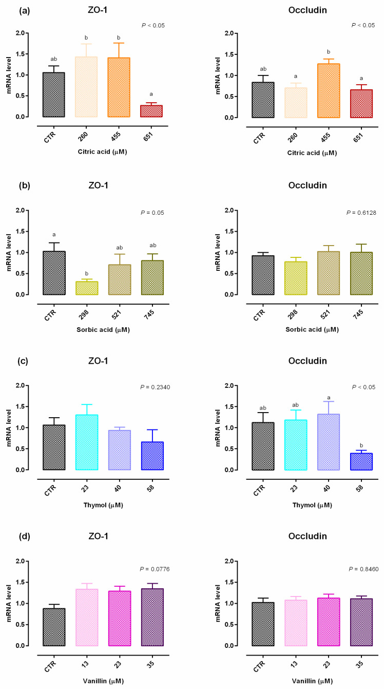
Figure 3.
Gene expression in Caco-2 cells after 15 days of treatment with single OA and NIC. Values are least square means (n = 6) ± SEM represented by vertical bars. A modification of the 2–ΔΔCT method [16] was used to analyze the relative expression (fold changes), calculated relative to the control group. Means with different letters indicate statistical significance with p < 0.05 (a, b); means with at least one common letter are not significantly different (ab). (a) Cells treated with citric acid; CTR = control group; 260 = treated group with 260 μM of citric acid; 455 = treated group with 455 μM of citric acid; 651 = treated group with 651 μM of citric acid. (b) Cells treated with sorbic acid; CTR = control group; 298 = treated group with 298 μM of sorbic acid; 521 = treated group with 521 μM of sorbic acid; 745 = treated group with 745 μM of sorbic acid. (c) Cells treated with thymol; CTR = control group; 23 = treated group with 23 μM of thymol; 40 = treated group with 40 μM of thymol; 58 = treated group with 58 μM of thymol. (d) Cells treated with vanillin; CTR = control group; 13 = treated group with 13 μM of vanillin; 23 = treated group with 23 μM of vanillin; 35 = treated group with 35 μM of vanillin. ZO-1 = zonula occludens.

