
Fig. 2.
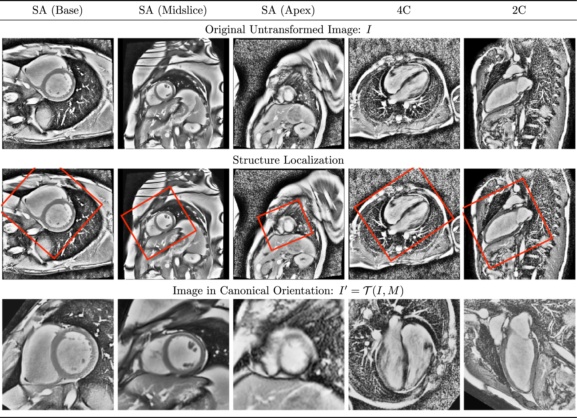
Orthogonal clinical views in canonical orientation Representative short axis (SA), four-chamber (4C), and two-chamber (2C) images are shown as acquired (top), and having undergone transformation into a canonical orientation (bottom). Consistent with common clinical practice, the heart is rotated such that in the SA views, the right ventricle appears on the (radiological) right side of the image, whereas in the 4C and 2C views, the long axis of the left ventricle is oriented vertically. The heart is also centered and scaled to fill 90% of the image. Note the heterogeneity in size, orientation, and appearance of the heart in the untransformed images, which contributes to the difficulty of segmentation.