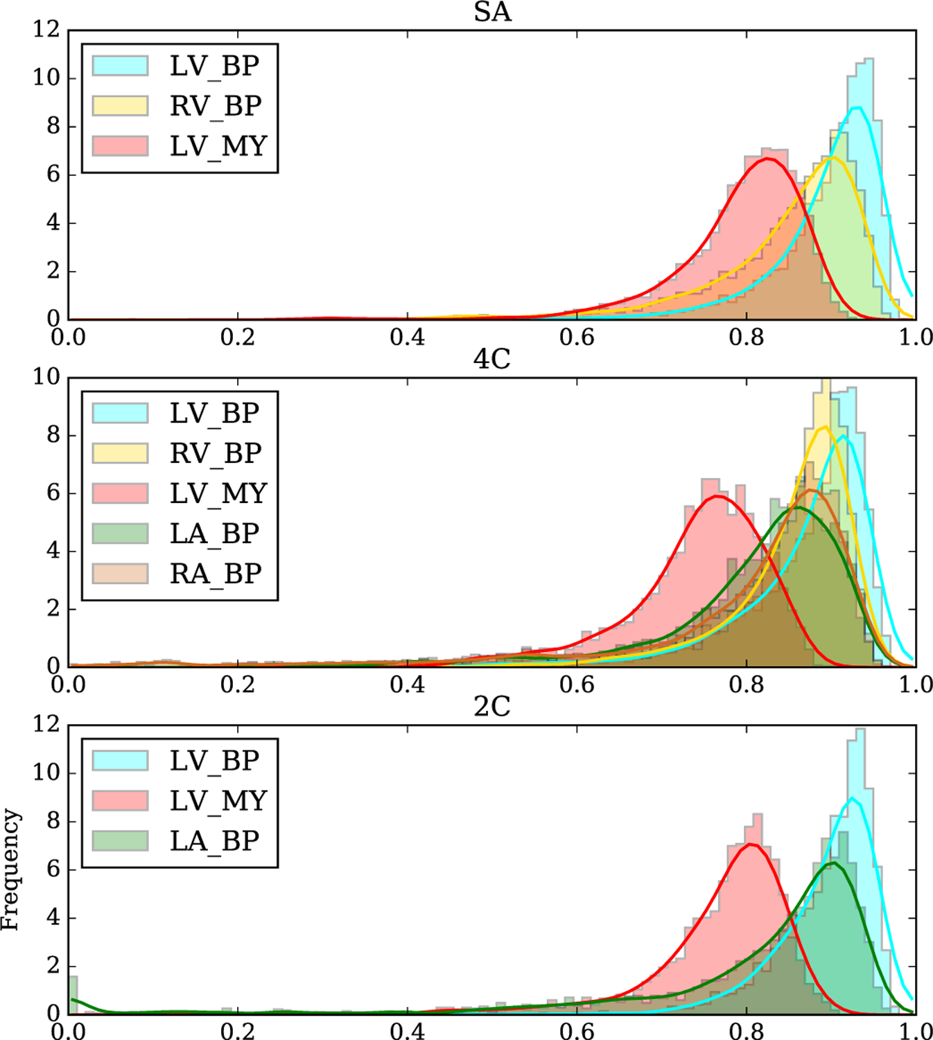
Fig. 7.

Histograms of IoU for each view and class. Performance relative to manual segmentation was highest in the SA view and lowest in the 4C view, though the differences are small. LV_BP: left ventricular blood pool; RV_BP: right ventricular blood pool; LV_MY: left ventricular myocardium; LA_BP: left atrial blood pool; RA_BP: right atrial blood pool.
