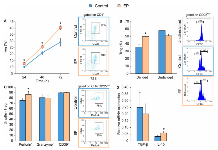
Figure 1.
The effect of ethyl pyruvate (EP) on Treg in vitro. CD4+CD25− T lymphocytes were stimulated for 24 h with a conventional Treg differentiation cocktail (control) and then additionally treated with 125 μM EP. (A) Treg (CD4+CD25highFoxP3+) proportion, at indicated time points after the addition of EP. Representative dot plots for the 72nd h are shown on the right-hand side. (B) Carboxyfluorescein succinimidyl ester (CFSE)-based proliferation of Treg (CD4+CD25high) was evaluated by flow cytometry (divided, Div. and undivided, Und. cells, with representative histogram plots shown on the right-hand side) 48 h after EP treatment. Unstimulated cells were stained with CFSE and served as the control for setting the threshold for undivided cells. (C) The proportion of Treg expressing perforin, granzyme or CD39, 72 h after EP treatment. Representative dot plots for perforin+ Treg are shown on the right-hand side. (D) mRNA expression of Tgf-β and Il-10 in cultures treated with or without EP. * p < 0.05 represents the significant difference of EP-treated cells in comparison to control cells.

