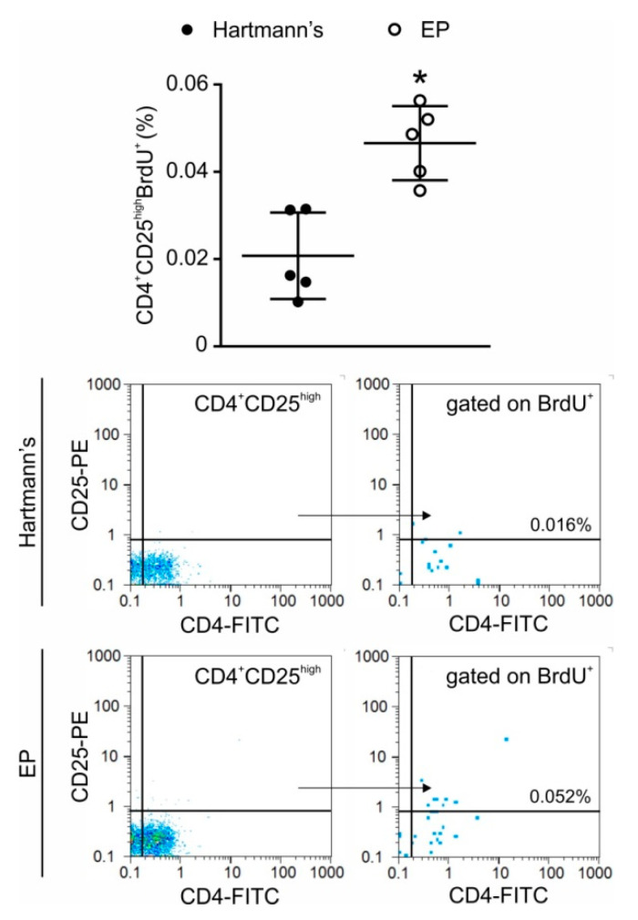
Figure 6.
Treg proliferation after intraperitoneal EP application. EP was administered twice a day for two days, while BrdU was applied intraperitoneally 24 h before ex vivo analysis. Treg (CD4+CD25high) proliferation was determined by flow cytometry. Representative dot plots and gating strategy are shown below the graph. * p < 0.05 represents the significant difference between EP-treated mice compared to vehicle-treated mice.

