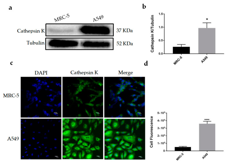
Figure 1.
Detection of the expression and location of Cathepsin K in cells. (a) Western blot (WB) analysis for Cathepsin K expression in MRC-5, A549 cells. Representative gel blots of Cathepsin K and Tubulin using specific antibodies. (b) Cathepsin K/Tubulin; (c) Cathepsin K immunofluorescence (IF) staining in MRC-5 and A549 cells. (d) The cell fluorescence intensity was calculated using Image J software (mean ± SEM, n ≥ 3, * p ≤ 0.05, ** p ≤ 0.01, *** p ≤ 0.001).

