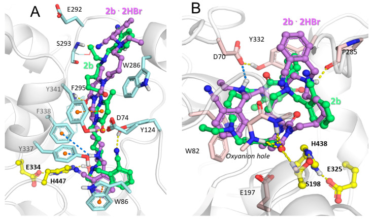
Figure 2.
Binding poses of 6-methyluracil 2b (carbon atoms are shown in green) and its charged counterpart 2b·2HBr (carbon atoms are shown in violet) inside active site gorges of (A) hAChE and (B) hBChE. Yellow, dashed lines represent hydrogen bonds, orange represents π–π stacking interactions, and blue lines indicate ionic and π–cation interactions. The catalytic triads of the enzymes are indicated using yellow carbon atoms.

