
Figure 8.
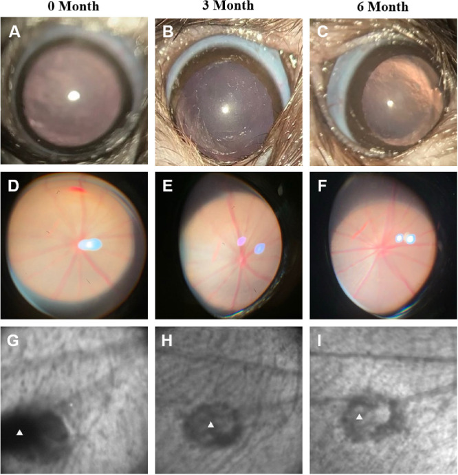
Representative slit-lamp and cSLO infrared examinations of eyes after DDS IVT injection. The first row shows slit-lamp photographs of the anterior chamber at 0, 3, and 6 months (A, B, and C) after DDS injection; the second row shows slit-lamp photographs of fundus at 0, 3, and 6 months (D, E, and F) after DDS injection; and the third row shows cSLO IR images of blank DDS (indicated by white triangles) at 0, 3, and 6 months (G, H, and I) after injection.