
Figure 9.
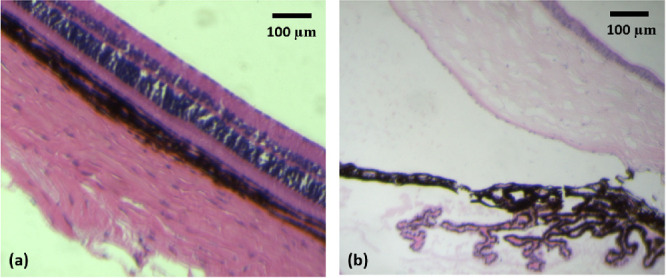
Representative end-point histopathology images of eyes after blank DDS IVT injection. (a) Bright-field microscopy of the retina 6 months after IVT injection of blank DDS. The ganglion cell layer is facing upward. (b) Bright-field microscopy of the anterior chamber 6 months after IVT injection of blank DDS. The iris tissue is facing upward.