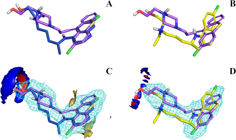
Figure 6.
Aligned structures of zuclopenthixol with R-HCQ (A) and S-HCQ (B). GRID fields intersection of both R-HCQ (C) and S-HCQ (D) with zuclopenthixol. Structures can be identified by the different color of carbon atoms: violet for zuclopenthixol, blue for R-HCQ and yellow for S-HCQ. GRID fields are colored as follows: red for hydrogen-bond donor, blue for hydrogen-bond acceptor, yellow for hydrophobic interaction. The size/shape field is shown as a light blue wireframe. Energy levels of the fields were tuned similarly for a better comparison across the figures.

