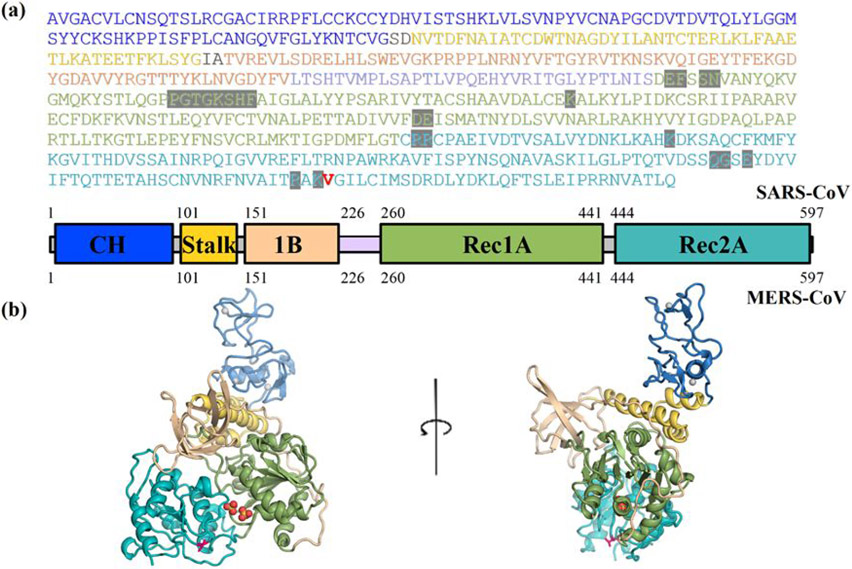
Figure 1. The SARS-CoV2 corona virus’s Nsp13 helicase structure.
(a) Sequence and domain structure of SARS-CoV2. The ATP binding site residues are highlighted in grey. The single residue V570 that is different between SARS-CoV2 and SARS (1570) in the Rec2A domain, is coloured red. The domain structure and colouring is shown below the sequence. (b) The (apo) SARS-CoV2 Nsp13 structural model (S2A) based on the I570V mutation of SARS Nsp13 (6JYT), coloured-by-domain, the V570 is show as red sticks. The domain structure and colouring scheme are the same as shown above.

