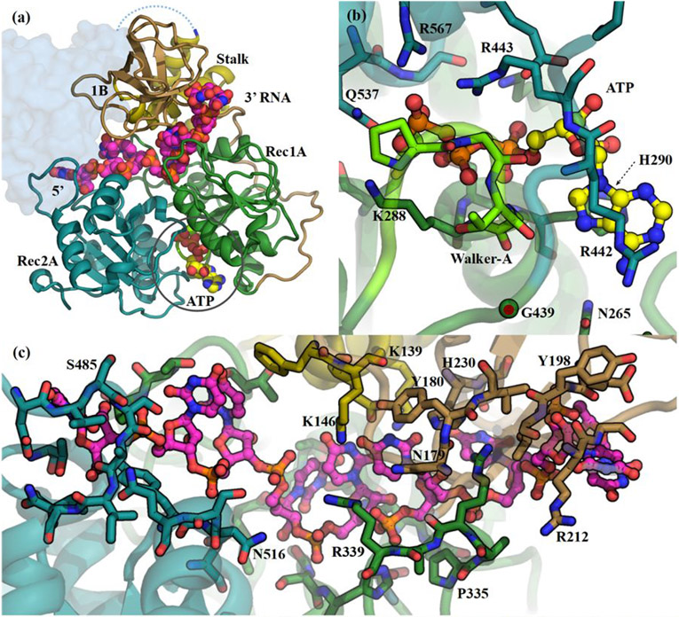
Figure 2. The SARS-CoV2 Nsp13:ATP:ssRNA complex model (S2C).
(a) The Domain organization in the S2C complex. Colouring is as in Figure 1. The ATP carbons are coloured yellow and the ssRNA carbons are magenta, both shown as spheres. The missing CH, Zn-binding, domain is shown as a pale-blue blob. (b) The ATP binding pocket showing specific interactions with Nsp13. View is from below the Rec2A (cyan) towards the Rec1A (green) domain. The Walker-A loop is bright green. (c) The ssRNA binds between the Stalk, Rec1A, and Rec2A domains. View is from the Stalk (yellow) towards the Rec1A (green) - 1B (tan) domain interface.

