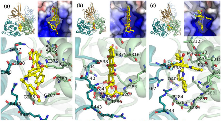
Figure 3. A Selection of apo-Nsp13 ATP binding site hits from virtual screening.
(a) Cepharanthine shown fit in the (S2A) SARS Nsp13 structure’s ATP binding site, (b) Idarubicin in M2B, (c) Nilotinib in M2B. Inset: Left: Overview of the inhibitor bound to the Nsp13. Right: The electrostatic surface in the active site (colour gradient: blue-to-red +/−5 V).

