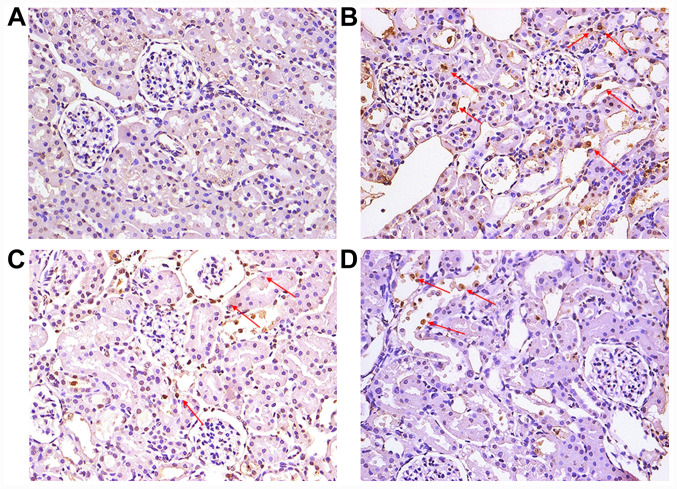
Figure 3.
Representative images of apoptotic characteristics of renal tubular epithelial cells. (A) normal control group. (B) model group. (C) human cord blood mononuclear cell group. (D) human umbilical cord-derived mesenchymal stem cell group. Arrows indicate representative apoptotic cells. Cells with brownish yellow granules in the nucleus were considered apoptotic. Magnification, x200.

