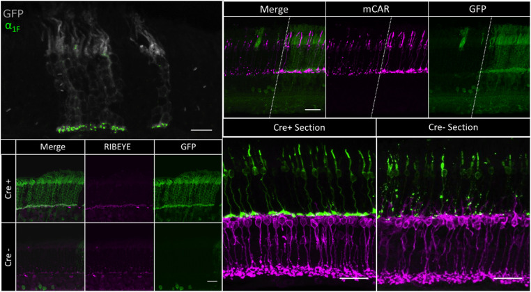
Figure 6.
(Upper Left) 60× cross-section of a recombined region of an iZEG:Cacna1f+;Pax6::Cre+;Cacna1fG305X mouse retina, immunolabeled for GFP (gray) and α1F (green). α1F-positive ribbon synapses can be observed in the OPL of GFP-positive columns (where the transgene is expressed and recombined by Cre). Scale bar: 10 µm. (Lower Left) 60× cross-section of recombined (top) and nonrecombined (bottom) regions of an iZEG:Cacna1f+;Pax6::Cre+;Cacna1fG305X mouse retina, immunolabeled for GFP (green) and RIBEYE (magenta). RIBEYE-positive elongated ribbon synapses can be observed in the OPL of the recombined region, whereas the nonrecombined region contains punctate RIBEYE immunolabeling scattered throughout the OPL and ONL, as previously described in the Cacna1f-KO retina.17 Scale bar: 10 µm. (Upper Right) 60x cross-section in a recombined region of an iZEG:Cacna1f+;Pax6::Cre+;Cacna1fG305 mouse retina, immunolabeled for GFP (green) and cone arrestin (magenta). Cone morphology is preserved in GFP-positive columns (where the transgene is expressed, and recombined by Cre), complete with pedicles, whereas cones in the non-GFP region exhibit signs of CSNB2A-related degeneration. Dotted lines separate recombined and non-recombined regions of the same retinal section. Scale bar = 10 µm (Bottom Right) 60x cross-sections of recombined (left) and non-recombined (right) regions of an iZEG: Cacna1f+; Pax6: Cre+; Cacna1fG305X mouse retina, immunolabeled for mCAR (green) and PKCα (magenta). Both cone axons and PKCα-positive bipolar cell dendrites terminate in a monolayer in the OPL in the recombined region, whereas cones exhibit axonal abnormalities and bipolar cells dendrites are sprouting into the ONL in the non-recombined region, as previously reported in the G305X mutant retina.17,20,22 Scale bar = 10 µm.

