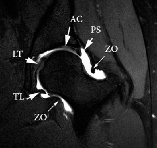
Figure 1.

Normal anatomy of the hip on magnetic resonance arthrogram (MRA). Coronal T1-weighted, fat-saturated MRA image of the hip demon- strating the zona orbicularis (ZO), transverse ligament (TL), ligamentum teres (LT), and peri-labral sulcus (PS). The articular cartilage (AC) of the acetabulum is seen to be continuous with the labrum
