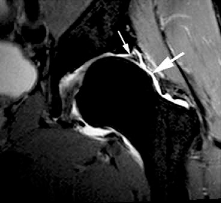
Figure 16.

Cam morphology and acetabular labral tear. Coronal T1W fat- saturated magnetic resonance arthrogram image showing a bone protu- berance at the lateral aspect of the femoral head and neck junction con- sistent with cam morphology (large arrow), and a chondrolabral junction intersubstance tear (small arrow). Note thinning of the articular cartilage at the acetabular margin
